- Research article
- Open access
- Published:
Determination of PCR efficiency in chelex-100 purified clinical samples and comparison of real-time quantitative PCR and conventional PCR for detection of Chlamydia pneumoniae
BMC Microbiology volume 2, Article number: 17 (2002)
Abstract
Background
Chlamydia pneumoniae infection has been detected by serological methods, but PCR is gaining more interest. A number of different PCR assays have been developed and some are used in combination with serology for diagnosis. Real-time PCR could be an attractive new PCR method; therefore it must be evaluated and compared to conventional PCR methods.
Results
We compared the performance of a newly developed real-time PCR with a conventional PCR method for detection of C. pneumoniae. The PCR methods were tested on reference samples containing C. pneumoniae DNA and on 136 nasopharyngeal samples from patients with a chronic cough. We found the same detection limit for the two methods and that clinical performance was equal for the real-time PCR and for the conventional PCR method, although only three samples tested positive. To investigate whether the low prevalence of C. pneumoniae among patients with a chronic cough was caused by suboptimal PCR efficiency in the samples, PCR efficiency was determined based on the real-time PCR. Seventeen of twenty randomly selected clinical samples had a similar PCR efficiency to samples containing pure genomic C. pneumoniae DNA.
Conclusions
These results indicate that the performance of real-time PCR is comparable to that of conventional PCR, but that needs to be confirmed further. Real-time PCR can be used to investigate the PCR efficiency which gives a rough estimate of how well the real-time PCR assay works in a specific sample type. Suboptimal PCR efficiency of PCR is not a likely explanation for the low positivity rate of C. pneumoniae in patients with a chronic cough.
Background
C. pneumoniae causes upper respiratory tract infections, and in some studies it accounts for 6–10% of community-acquired pneumonia [1]. Seroprevalence among adults is 40–70%, increasing with age, indicating that most people are exposed at least once and that reinfections are common [2]. C. pneumoniae is the microorganism that most commonly is associated with the inflammation seen in atherosclerosis [3].
C. pneumoniae infection has been detected by serological methods but PCR is currently viewed as an advantageous alternative since it detects the presence of the DNA of the organism. This allows for an early and clinically relevant diagnosis in contrast to the detection of C. pneumoniae specific antibodies that develop late in the course of the infection. Today, there is no accepted gold standard for PCR detection of C. pneumoniae, but as a start, guidelines for standardising C. pneumoniae assays have been published [4]. Most laboratories use in-house PCR assays and DNA purification procedures. Real-time quantitative PCR in the Lightcycler is a new option among the many PCR assays and it may become a method of choice as it is fast and provides a quantitative measure. This is advantageous in a clinical setting because it allows fast diagnosis and thus fast treatment with relevant antibiotics. Furthermore, the quantitative measure could possibly be used to assess the response to treatment or to assess the state of infection; active infection versus chronic infection.
The lack of standardisation of PCR methods has been investigated in three recent studies [5–7]. These studies found strikingly different results when comparing several PCR methods. The inconsistent results were explained by the use of different DNA extraction methods, different thermal cyclers and differences in number of and/or homogeneity of replicates. Contradicting C. pneumoniae PCR results have also been explained by the fact that C. pneumoniae is often present in very low numbers because the infections tend to be chronic and low-grade [8]. Even though most assays have very good detection limits the fact that they are often used at DNA concentrations near their detection limit can pose problems. At low DNA concentrations detection/non-detection is more susceptible to inhibitors because inhibition adds further to the statistical uncertainty already present at the detection limit (i.e. it is only possible to detect 1 copy of DNA in a fraction of experiments, because the particle distribution is expected to follow Poisson statistics). Therefore, it is necessary to test PCR methods for their performance in different situations and sample types.
The aim of this study was to compare a newly developed real-time quantitative PCR in the Lightcycler for detection of C. pneumoniae[9] with an established conventional PCR assay used in another laboratory [10, 11] to see whether results obtained with the two methods correlate. In the real-time PCR the pmp4 gene is target and in the conventional PCR it is the 16S rRNA gene. PCR's were performed in the laboratories where they were developed. The limit of detection and performance on clinical specimens were compared and advantages and disadvantages of the methods are discussed. PCR efficiency was determined by the real-time PCR method in a random selection of the clinical samples to test whether inhibitors were present.
Results and discussion
Reference samples: comparison of the two PCR methods
C. pneumoniae-infected HeLa cells were Proteinase K-treated and a serial four-fold dilution series was prepared with concentrations near and past the detection limit for both PCR methods used. These reference samples were tested with a real-time PCR and a conventional PCR both specific for C. pneumoniae (see table 1 for sample volumes). The results obtained from the reference samples with the two PCR methods are shown in table 2. Samples were blinded prior to analysis, to avoid experimentator-induced bias. Both PCR methods correctly identified the five first samples in the dilution series, and no false positives were observed. In the conventional PCR (method 2) it is an advantage that a higher template volume can be used, which might make it possible to detect lower C. pneumoniae concentrations. This is not possible for the real-time PCR method (method 1) as the capillaries limit the reaction volume to 20 μl. In conclusion, the two PCR methods had the same limit of detection. We avoided using inclusion-forming units (IFU) as a measure of sensitivity because it is probably not very informative as different culture systems may vary considerably and because the determination procedure, which involves cultivation and counting under a microscope, can be operator dependent [8]. Measuring DNA concentration with a spectrometer is less operator dependent and is therefore probably easier to compare between laboratories. Furthermore, IFU only measures the number of viable EB in a preparation, it does not account for RB and dead EB, which also contain genomic DNA amplifiable by PCR.
Determination of PCR efficiency in the reference and standard samples with real-time PCR
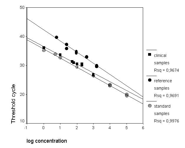
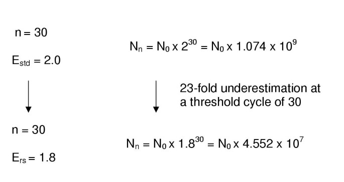
When performing quantitative real-time PCR, it is assumed that the PCR efficiency of the standard samples is the same as that of the unknown samples. This is the basis for the calculations made. Therefore, it is important to determine the efficiency in both standards samples and unknown samples. PCR efficiency depends e.g. on inhibition in the sample, how well primers and probes are designed, and how well the PCR conditions are optimised. PCR efficiency was determined in the reference samples described above and in standard samples. The standard samples were a 10-fold dilution series of purified genomic C. pneumoniae DNA with known concentrations. Threshold cycles obtained with the real-time PCR for duplicates of the reference and standard samples were plotted against log to the concentration of the samples (log to the concentration: arbitrary numbers with four-fold difference for the reference samples, as concentrations were unknown) (fig. 1). Two straight lines were drawn and the slopes were determined by linear regression to be -3.848 (standard error = 0.259) for the reference samples and -3.144 (standard error = 0.065) for the standard samples. The two slopes were found to be significantly different (z = 2.64, P = 0.008). Efficiency is derived from the idealized function for the amount of PCR product formed: N = N0 × En, where N is number of amplified molecules, N0 is the initial number of molecules, n is the number of amplification cycles and E is the efficiency which is ideally 2. The standard curves are derived from the function described above: n = -(1/log E) * log N0 + (log N/log E). Therefore, the slope of the line equals -(1/ log E) and the efficiency can be calculated from the slope. From the slopes the efficiency of the real-time PCR in the reference samples was determined to be 1.82 and in the standard samples to be 2.08. The fact that the efficiency is a little larger than 2 is probably caused by the linear regression analysis which does not take into account that the efficiency can maximally be 2. Because of the difference in efficiency between reference and standard samples an inaccuracy in the determined concentration of the reference samples is present. As the efficiency in the reference samples is lower, the concentration determined on basis of the standard samples will be an underestimation. This is seen in table 2, as it would be highly unlikely to be able to determine the lowest concentration 0.05 copies/μl if it was not an underestimation caused by different efficiencies. The underestimation factor can be estimated for each cycle number. At cycle number 30 and with the described difference in efficiency the concentration would be underestimated 23-fold (see fig. 2). Differences in efficiency of PCR can be caused by several factors e.g. inhibitors and storage of the sample, but different batches of primers, probes and enzymes might also influence the efficiency. The DNA for the reference samples was released by Proteinase K treatment only with no subsequent purification; therefore inhibitors might have been present. Furthermore, as a larger volume of reference sample (6.8 μl) than standard sample (2 μl) was used, this could increase inhibition. On the other hand, the amplification efficiency appeared to be identical for all dilutions as indicated by the regression line, so inhibition from the sample preparation might not be the explanation; the reference samples had been stored in polypropylene tubes for more than a month before analysis, and as DNA binds to polypropylene [12], this could also affect the efficiency observed as this effect alters the DNA concentrations especially in the dilute samples [12].
Determination of the efficiency for the standard, reference and clinical samples. The threshold cycle is the cycle number at which the fluorescence curve for a given sample crosses the user-defined noise band. Log to the concentration for the reference samples is set as log to arbitrary numbers with four-fold difference as the reference samples are four-fold serial dilutions. The actual concentration is not needed when determining the efficiency as it depends on the slope of the line only.
Effect of difference in efficiency between standard samples and samples. This figure illustrates the error in determination of concentration by real-time PCR at cycle no. 30 when the efficiencies differ between standard samples and samples. Number of cycles: n, Efficiency standard samples: Estd, efficiency reference samples: Ers, Number of amplicons at cycle n: Nn, Number of amplicons at cycle zero: N0. When the two equations are divided by eachother it is seen that there is an 23-fold underestimation of the concentration when Estd = 2.0 and Ers = 1.8. From this calculation it is also evident that at higher cycle number the error in concentration determination is larger.
Clinical samples: comparison of the two PCR methods
Nasopharyngeal aspirates were previously obtained from patients with chronic cough [9] and DNA was released with Chelex 100. A selection of 136 clinical samples was used. The results obtained from the clinical samples with the two PCR methods are shown in table 3. Method 1 found three of 136 samples to be positive and method 2 found the same three samples to be positive, and both methods found the rest of the samples negative. Therefore, there seems to be good concordance between the two PCR methods, but more samples are needed to confirm this. The three patients found positive for C. pneumoniae by PCR by both methods were C. pneumoniae IgM positive. No PCR negative patients were found to be IgM positive. Therefore, in this study, PCR correlates with the IgM positivity, meaning that even a small amount of C. pneumoniae DNA in samples (table 3) may reflect an infection capable of initiating an immune response. This could be because the clinical samples are from patients with chronic and thereby long-lasting cough. The infection they have does probably not display high bacterial loads, but more likely it is an infection with a low load of C. pneumoniae. In order to assess whether the positivity rate was underestimated due to suboptimal efficiency of the real-time PCR, we performed efficiency analyses on the three positive clinical specimens and a random selection of negative clinical samples. Even though there is concordance between PCR and IgM results, it is believed that IgM may not be present if it is a reinfection [13]. Therefore it is possible that PCR is positive when IgM is not.
Spiked clinical samples: determination of PCR efficiency by real-time PCR
The efficiency of real-time PCR in the positive clinical sample with the highest C. pneumoniae DNA concentration (no. 133) was determined as described above and compared to the efficiency for the standard samples (fig. 1). A dilution series was prepared of sample 133 and the threshold cycles determined for duplicates (dilutions: 5, 10, 15, 20, 100 and 1000 times). The slope of the line (fig. 1) was -3.237 (standard error = 0.158) indicating an efficiency of 2.0. The slope for clinical sample line was not significantly different from the slope for the standard samples line (slope for the standard: -3.144 and standard error: 0.065, t-test: z = 0.54, P = 0.59).
In table 4 slopes and standard errors for dilution series of negative samples spiked with 103 copies/μl C. pneumoniae DNA and the two samples (samples no. 1 and 60) with low C. pneumoniae DNA concentration are shown. Two-fold dilution series were prepared for each sample. The slopes for the spiked samples were compared to the slope of the standard samples from the same run. P-values for a t-test testing whether slopes differ significantly are shown. Three (sample no's. 101, 131 and 160) of the 20 samples have slopes significantly different from the slope of the corresponding standard and none of these were C. pneumoniae positive when not spiked. This means that the efficiency differs significantly and quantification of these samples is not accurate. It should be noted though, that for the samples 1, 60 and 133 (the three C. pneumoniae positive samples) there are no signs of inhibition. Therefore, quantification of the positive samples is probably reasonably accurate.
For the remaining 17 samples the slopes were not significantly different indicating that the efficiencies should match satisfyingly between standard samples and spiked clinical samples. Hence, the PCR conditions seem to be similar in the clinical and standard samples, therefore detection and quantification is possible. Consequently, it is probably unlikely that larger numbers of positive samples were missed because of suboptimal PCR efficiency. If PCR efficiency is in part influenced by inhibitors, our results indicate that Chelex release of DNA from nasopharyngeal aspirates is sufficient in most samples. However, if the nasopharyngeal aspirates are contaminated with e.g. blood, which contain many PCR inhibitors, further purification might be needed.
It should be noted that even though slopes are not significantly different, there might still be differences in efficiencies, as the t-test allows for some variation. In table 4 it is also seen that some of the efficiencies are larger than 2, which should not be possible according to PCR theory. Some of the samples have efficiencies very much larger than 2 (samples 131 and 160) and these were also found to differ significantly from efficiencies of the standards. Substances in the samples that interfere with the PCR probably cause this. In the samples with efficiency just slightly higher than 2, it is probably caused by the fact that the slope is determined by linear regression. The linear regression does not take into account that the slope can maximally be -3.32, which corresponds to an efficiency of 2.0. Furthermore, it is notable that slopes for the standard samples vary from -3.266 to -3.716 (E varies from 1.86–2.01). Many things may cause this variation. First of all, variations in preparation of the standard dilution series is difficult to avoid because of pipetting variations, but also because DNA molecules do not behave like smaller molecules in solution. DNA is a large molecule and it is therefore likely to make intramolecular (electrostatic) interactions and also interactions with other molecules. These interactions are hard to predict as they depend on 1) which molecules are present and their concentration in the solution, 2) temperature 3) length and integrity of the DNA molecules. These phenomena probably contribute to the variation. This emphasizes that when a quantitative PCR result is presented it should be interpreted with care and always in the context of the experimental setup. When comparing quantitative measures determined in this way, it should probably be recommended to run the samples in the same run, to run at least two replicates to obtain a mean value or to include internal amplification controls. Even then the results should be interpreted with care. It is possible to detect relatively small differences between sample concentrations with quantitative real-time PCR, but the exact factor of difference between samples should be interpreted very cautiously. We have observed this previously [9] with our standard dilution series; in most runs it is actually possible to distinguish between e.g. 10 copies/μl and 5 copies/μl standard samples, but the factor of difference, if exact quantitative measures are compared, can vary substantially. Furthermore, the fact that the crossing points determined for the unknown samples are proportional to the log of the concentration and not directly to the concentration is also a major error factor because invlog enlarges the errors when concentration is calculated.
In practice it would not be feasible to test the PCR efficiency for every sample and in every run, because of the added time and costs. Therefore, it should be done when setting up a new real-time PCR assay and DNA purification method for a new clinical sample type. In our opinion, based on this study, the efficiency can provide a rough estimate for how well the PCR assay performs in a specific sample type, but small differences in efficiency should be interpreted cautiously because as described above many error factors are involved in the determination of this parameter.
Analysing replicates could be relevant for nasopharyngeal aspirates as C. pneumoniae might be present in very low numbers. The reason could be that cells in the nasopharyngeal aspirates stem from the superficial epithelium and C. pneumoniae might be present in larger numbers in deeper cell layers [13]. The disadvantages of analysing replicate samples are that it adds extra costs, time and strict control of contamination. As C. pneumoniae tends to be present in low numbers especially in non-acute patients as in this study, it might be more feasible to attempt concentrating samples before or after DNA extraction [7, 14] or to improve sampling methods.
Conclusions
In this study sensitivity and clinical performance seems to be the same for conventional PCR and real-time PCR, but this needs to be confirmed by analysis of more clinical samples. We extended the investigation of clinical performance with determination of the PCR efficiency in the samples. The efficiencies determined by real-time PCR in clinical samples were in 17/20 cases determined not to be significantly different from efficiencies of the standards. This means that suboptimal PCR efficiency was probably not the explanation for the low positivity rate of C. pneumoniae in the nasopharyngeal aspirates. The efficiency may be used as a rough estimate for how well real-time PCR works in a given sample type.
Materials and methods
PCR methods
Method 1
Real-time quantitative PCR was performed in a Lightcycler instrument as described previously [9]. Briefly, the real-time PCR amplifies a 140 bp PCR product from the pmp4 gene of C. pneumoniae. The PCR fragment is detected with a fluorescent probe set using fluorescence resonance energy transfer. The pmp4 gene encodes a membrane protein from the polymorphic membrane protein (Pmp) family, which contain 21 proteins [15]. Each gene is present in one copy on the genome, homologs are found in C. psittaci and C. trachomatis, but the sequences are not very similar on DNA level, therefore it is possible to design species specific primers and probes. Specificity was assessed by database searches with the primer and probe sequences and pmp4 gene sequences were compared for the known genome sequences of C. pneumoniae. Primer and probe sequences were found to be specific for C. pneumoniae and the pmp4 gene sequences for the known isolates were identical [9]. Real-time PCR was performed in 20 μl consisting of: 0.5 μm of each primer, 0.2 μm of each probe, 5 mM MgCl2, 2.0 μl Faststart DNA Master Hybridisation Probes (Roche) and 2 or 6.8 μl template. The mixture was loaded into glass capillary tubes and cycling performed as previously described [9] The PCR was performed with 6.8 μl template DNA for analysis of the reference samples and 2 μl for analysis of the clinical samples and standard samples (see below for definition of samples). For easy comparison sample volumes are shown in table 1.
Standard samples
C. pneumoniae EB was purified by gradient centrifugation as previously described [16] and genomic DNA was purified with the Genomic-tip system from Qiagen following instructions from the manufacturer. DNA concentration and purity were analysed by agarose gel electrophoresis and measurement of OD at 260 and 280 nm. A ten-fold standard dilution series of purified C. pneumoniae genomic DNA with known concentration was used for quantification, concentrations ranged between 105 and 1 copies/μl [9]. The Lightcycler software determined the threshold cycles for the standard samples and a standard curve was generated. The threshold cycle for unknown samples was determined for two replicates and concentrations were calculated from the standard curve also using the Lightcycler software. A negative control containing all reagents except template DNA was included in all runs. A sample was considered to be positive when the fluorescence curve was sigmoidal and reached fluorescence levels above the fluorescence threshold set in the Ligthcycler software during analysis.
Method 2
A multiplex PCR with three primer sets was used which amplify fragments from the 16S rRNA gene of the three Chlamydia species. The primer pair specific for C. trachomatis, which amplifies a 240 bp product, was designed by Pollard et al. [17], from these primers analogues were designed for the other Chlamydia species [11]. An internal amplification control was included in order to detect inhibition or suboptimal reaction conditions [11, 17, 18]. Amplicons were visualised with ethidium bromide and agarose gel electrophoresis. Positive results were confirmed with a C. pneumoniae specific PCR with primers detecting a 463 bp fragment of the 16S rDNA [19]. For analysis of the reference samples 10 μl template DNA was used. For analysis of the clinical samples 10 μl was used. If inhibition occurred, the assay was repeated with 5 μl or 2 μl of the sample. Samples volumes are shown in table 1 for both PCR methods.
For both PCR methods precautions were taken to avoid contamination. Filter pipette tips were used and reagents were mixed in rooms separated from rooms where C. pneumoniae DNA from culturing, DNA purification etc. was present.
Reference samples
C. pneumoniae was cultivated in HeLa cells as previously described [20]. Cells were scraped off with a rubber policeman, centrifuged and resuspended in TE-buffer. Proteinase K was added and the mixture was incubated for one hour at 55°C. Proteinase K was inactivated by incubation at 95°C for 10 min. A serial four-fold dilution series was prepared in TE-buffer with 10 dilutions ensuring a wide range of concentrations. Samples with C. pneumoniae DNA concentrations near the detection limit should therefore be present for the methods tested. In addition, two negative controls containing TE-buffer only were included. The two PCR methods were tested on the reference samples and their relative sensitivities were obtained.
Clinical samples and spiked clinical samples for efficiency determination
Clinical samples
Nasopharyngeal aspirates were previously obtained from patients with chronic cough from the Dept. of Pulmonary Medicine at Aarhus University Hospital, Denmark. Inclusion criteria were age >15 yrs., a cough period of 2–12 weeks, a normal chest radiograph, normal spirometry and absence of chronic cardiopulmonary disease [10]. For this study a selection of 136 samples was used. DNA was released from the nasopharyngeal aspirates by vortexing with 20% w/v Chelex 100 slurry followed by boiling of the suspensions for 10 min; then centrifuged at 20,000 × g for 10 min. [10]. Chelex-100 is an ion-exchange resin that scavenges multivalent metal ions. PCR methods 1 and 2 were tested on these samples.
Spiked clinical samples
The real-time PCR was used for determination of PCR efficiency in a selection of clinical samples in the following way. For negative samples and samples with low C. pneumoniae DNA concentration spiking with 103 copies/μl C. pneumoniae was done. Afterwards a 2-fold dilution series with 8 dilutions was done in water with 10 μg/ml yeast RNA as carrier nucleic acid [9]. For each of the dilutions we measured threshold cycles and plotted them towards log to relative concentrations of the dilutions. The slope was determined and compared to the slope of the standard samples from the same run.
References
Grayston JT: Infections caused by Chlamydia pneumoniae strain TWAR. Clin Infect Dis. 1992, 15 (5): 757-761.
Peeling RW, Brunham RC: Chlamydiae as pathogens: new species and new issues. Emerg Infect Dis. 1996, 2 (4): 307-319.
Saikku P: Chlamydia pneumoniae in atherosclerosis. J Intern Med. 2000, 247 (3): 391-396. 10.1046/j.1365-2796.2000.00659.x.
Dowell SF, Peeling RW, Boman J, Carlone GM, Fields BS, Guarner J, Hammerschlag MR, Jackson LA, Kuo CC, Maass M, Messmer TO, Talkington DF, Tondella ML, Zaki SR: C. pneumoniae Workshop Participants. Standardizing Chlamydia pneumoniae assays: recommendations from the Centers for Disease Control and Prevention (USA) and the Laboratory Centre for Disease Control (Canada). Clin Infect Dis. 2001, 33 (4): 492-503. 10.1086/322632.
Mahony JB, Chong S, Coombes BK, Smieja M, Petrich A: Analytical sensitivity, reproducibility of results, and clinical performance of five PCR assays for detecting Chlamydia pneumoniae DNA in peripheral blood mononuclear cells. J Clin Microbiol. 2000, 38 (7): 2622-2627.
Apfalter P, Blasi F, Boman J, Gaydos CA, Kundi M, Maass M, Makristathis A, Meijer A, Nadrchal R, Persson K, Rotter ML, Tong CY, Stanek G, Hirschlm AM: Multicenter Comparison Trial of DNA Extraction Methods and PCR Assays for Detection of Chlamydia pneumoniae in Endarterectomy Specimens. J Clin Microbiol. 2001, 39 (2): 519-524. 10.1128/JCM.39.2.519-524.2001.
Smieja M, Mahony JB, Goldsmith CH, Chong S, Petrich A, Chernesky M: Replicate PCR Testing and Probit Analysis for Detection and Quantitation of Chlamydia pneumoniae in Clinical Specimens. J Clin Microbiol. 2001, 39 (5): 1796-1801. 10.1128/JCM.39.5.1796-1801.2001.
Boman J, Gaydos CA, Quinn TC: Molecular diagnosis of Chlamydia pneumoniae infection. J Clin Microbiol. 1999, 37 (12): 3791-3799.
Mygind T, Birkelund S, Falk E, Christiansen G: Evaluation of real-time quantitative PCR for identification and quantification of Chlamydia pneumoniae by comparison with immunohistochemistry. J Microbiol Meth. 2001, 46: 241-251. 10.1016/S0167-7012(01)00282-2.
Birkebæk NH, Jensen JS, Seefeldt T, Degn J, Huniche B, Andersen PL, Østergaard L: Chlamydia pneumoniae infection in adults with chronic cough compared with healthy blood donors. Eur Respir J. 2000, 16 (1): 108-111. 10.1034/j.1399-3003.2000.16a19.x.
Storgaard M, Østergaard L, Jensen JS, Farholt S, Larsen K, Ovesen T, Nødgaard H, Andersen PL: Chlamydia pneumoniae in children with otitis media. Clin Infect Dis. 1997, 25 (5): 1090-1093.
Belotserkovskii BP, Johnston BH: Denaturation and association of DNA sequences by certain polypropylene surfaces. Anal Biochem. 1997, 251 (2): 251-262. 10.1006/abio.1997.2249.
Kauppinen M, Saikku P: Pneumonia due to Chlamydia pneumoniae: prevalence, clinical features, diagnosis, and treatment. Clin Infect Dis. 1995, 21 (Suppl. 3): S244-S252.
Maass M, Jahn J, Gieffers J, Dalhoff K, Katus HA, Solbach W: Detection of Chlamydia pneumoniae within peripheral blood monocytes of patients with unstable angina or myocardial infarction. J Infect Dis. 2000, 181 (Suppl 3): S449-S451. 10.1086/315610.
Kalman S, Mitchell W, Marathe R, Lammel C, Fan J, Hyman RW, Olinger L, Grimwood J, Davis RW, Stephens RS: Comparative genomes of Chlamydia pneumoniae and C. trachomatis. Nat Genet. 1999, 21 (4): 385-389. 10.1038/7716.
Knudsen K, Madsen AS, Mygind P, Christiansen G, Birkelund S: Identification of two novel genes encoding 97- to 99-kilodalton outer membrane proteins of Chlamydia pneumoniae. Infect Immun. 1999, 67 (1): 375-383.
Pollard DR, Tyler SD, Ng CW, Rozee KR: A polymerase chain reaction (PCR) protocol for the specific detection of Chlamydia spp. Mol Cell Probes. 1989, 3 (4): 383-389.
Tarp B, Jensen JS, Østergaard L, Andersen PL: Search for agents causing atypical pneumonia in HIV-positive patients by inhibitor-controlled PCR assays. Eur Respir J. 1999, 13 (1): 175-179. 10.1034/j.1399-3003.1999.13a32.x.
Gaydos CA, Quinn TC, Eiden JJ: Identification of Chlamydia pneumoniae by DNA amplification of the 16S rRNA gene. J Clin Microbiol. 1992, 30 (4): 796-800.
Clausen JD, Christiansen G, Holst HU, Birkelund S: Chlamydia trachomatis utilizes the host cell microtubule network during early events of infection. Mol Microbiol. 1997, 25 (3): 441-449. 10.1046/j.1365-2958.1997.4591832.x.
Acknowledgements
The Danish Medical Research Council supported the research (grants no. 9900750 and 9700659). We thank Inger Andersen, Karin Skovgaard Sørensen, Birthe Dohn and Jonna Guldberg for technical assistance and Lisbet Wellejus Pedersen for linguistic assistance
Author information
Authors and Affiliations
Corresponding author
Additional information
Authors' contributions
Author T.M. carried out the real-time PCR experiments and the statistical analysis of the data and drafted the manuscript. Author S.B. participated in design and coordination of the study. Author N.H.B collected the clinical samples. Author L.Ø. participated in design and coordination of the study and provided reference samples. Author J.S.J. carried out the conventional PCR experiments. Author G. C. participated in design and coordination of the study.
Authors’ original submitted files for images
Below are the links to the authors’ original submitted files for images.
Rights and permissions
About this article
Cite this article
Mygind, T., Birkelund, S., Birkebæk, N.H. et al. Determination of PCR efficiency in chelex-100 purified clinical samples and comparison of real-time quantitative PCR and conventional PCR for detection of Chlamydia pneumoniae. BMC Microbiol 2, 17 (2002). https://doi.org/10.1186/1471-2180-2-17
Received:
Accepted:
Published:
DOI: https://doi.org/10.1186/1471-2180-2-17

